Tytuł pozycji:
Kartoteka oceny histopatologicznej chorób układu nerwowego (1963) - opis nr 184/63
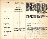
Histological diagnosis: Atrophia cerebri. Arteriosclerosis cerebri.Autopsy examination of 71-year-old patient was performed. Neuropathological evaluation in light microscopy was based on brain paraffin sections stained with Hematoxylin-eosin and van Gieson method and frozen section s stain with Bielschowsky method.In the cortex, cellular desolations were observed, obliterating in parts its layered structure. Nerve cells were often steatotic, some showed features of chronic disease. The changes were most pronounced in the third and fifth layer and perivascularly. Large atherosclerotic lesions were present. The vessels, especially in the basal ganglia and white matter, presented changes typical of vitrification and fibrosis, fibrous changes were observed in the cortex. Lymphocyte-like cells, macrophages with hemosiderin and pseudocalcium salts were seen near some vessels. Besides, there were features of congestion and edema. In the meninges, single macrophages were present in some areas. No senile plaques were found in the cortex or basal ganglia.
W korze opustoszenia komórkowe zacierające odcinkami warstwową budowę.Komórki nerwowe często stłuszczałe,niektóre wykazują cechy schorzenia przewlekłego. Największe nasilenie zmian występuje w III i V-tej warstwie oraz przynaczyniowo. Duże zmiany miażdżycowe. Naczynia zwłaszcza zwojów podstawy i istoty białaj wykazują zmiany typowe dla szkliwienia i włóknienia /III i IV0/. w kórze zmiany włókniste. Przy niektórych naczyniach k. limfocytopodobne, makrofagi z hemosyderyną, sole pseudo-wapnia. Poza tym cechy przekrwienia i obrzęku. W oponach gdzieniegdzie pojedyńcze makrofagi. Plak starczych w korze ani w zwojach podstawy w barwieniach Bielschowskyego nie stwierdziłam.
Clinical, anatomical and histological diagnosis
Rozpoznanie kliniczne, anatomiczne i histologiczne

